


1.1.3 The Heart and Electrocardiography ECG
Intrinsic Conduction System of the Heart
The heart is the main organ in the cardiovascular system. When the heart contracts its walls, it pumps the blood through the blood vessels; transporting nutrients, oxygen and waste to and from body cells. These contractions result from electrical depolarisation waves that travel through the cardiac muscle cells, causing them to beat rhythmically as a single unit. This ability does not depend on impulses from the nervous system.
The cells in charge of the intrinsic conduction of the heart are the cardiac pacemaker cells. They guarantee that the heart muscle depolarises and beats in a sequential manner, from atria to ventricles. The figure below depicts the impulses traveling through the heart (from 1 to 5) following the yellow indications.
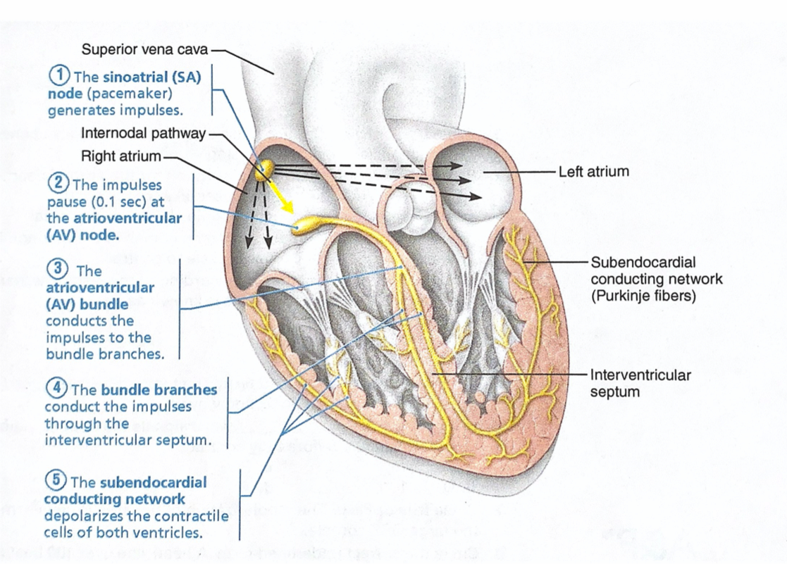
Source: E. Marieb & L. Smith. “Human Anatomy & Physiology Laboratory Manual, 10th Edition“. 2014
The heart has four hollow chambers— two atria (in singular is called atrium) and two ventricles .
- The Atria are primarily receiving chambers. As a rule, they are not important in the pumping activity of the heart. Instead, they assist with filling the ventricles. Blood flows into the atria under low pressure from the veins of the body and then continues on to fill the ventricles.
- The Ventricles are the thick-walled discharging chambers, or actual pumps of the heart. When they contract, blood is propelled out of the heart into circulation.
Electrocardiography ECG
An electrocardiograph is an instrument that detects and records the electrical currents on the body’s surface, generated by the impulses of the heart. The graphic recording of the heart’s electrical impulses is known as electrocardiogram or ECG, and its characterised by the following components:
- Deflection Waves: they correspond to the depolarisation and repolarization of the heart’s chambers. A typical ECG has 3, the P wave, the QRS complex and the T wave.
- Segment: is a region between two waves. For example, the T-P segment.
- Interval: which is the region between a segment and one or more waves. For example, the Q-T interval covers the QRS complex, S-T segment and T wave.
Abnormalities found in the waves of an ECG are useful to detect heart anomalies, like a heart attack. The relationship between the deflection waves of an ECG, and the sequence of repolarization and depolarisation is described in the image bellow.
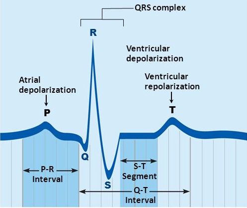
Source: E. Marieb & L. Smith. “Human Anatomy & Physiology Laboratory Manual, 10th Edition”. 2014
ECG Recording
An ECG recording is performed when an arrhythmia or any other heart anomalies are suspected. It is a painless and non-invasive procedure. Electrodes are attached to the body, usually on the arms, legs and across the chest. They detect the small electrical impulses as described earlier.
For diagnostic purposes, the standard is to perform a 12-lead ECG, where there are 12 leads calculated using 10 electrodes. Four are placed on each arm/leg and six on the chest, close to the heart. The 12 leads provide a comprehensive picture to the doctor of the heart’s electrical activity. The Lead I, II and III are bipolar since they measure the voltage difference between different limbs; while the other 9 leads are unipolar.
If you want to learn more about the ECG lead placement and the variations in the lead placement we recommend the following link as additional material: